Draw And Label Sponges : Sponges (Porphyria) at Moravian College - StudyBlue / List 4 ways sponges defend themselves.. The difference between this and the mechanisms of other animals. The sponge to draw in ambient water through small inhalant pores (ostia) and ilter out microscopic food particles. Explain the process of reproduction in the sponge: Scattered among the pinacoderm are the ostia that allow entry of water into the body of the sponge. Hermaphroditic, gamete, spawn, planktonic larvae.
Label the collection tubes at the bedside or drawing area. A collagenous matrix (the mesohyl) ills the space between the canals and chambers, harbouring Explain how a stinging cell in a cnidarian fires. The instructions included specific information about sponge anatomy to be colored on diagram. This problem has been solved!
Overview of Sponges from web.augsburg.edu Asexual methods of reproduction include: How to draw hydrahow to draw hydra, how to draw hydraulic circuit diagram, how to draw hydra step by step, how to draw hydra easy, how to draw hydra in zoolo. Spongin, spicules, ostia, choanocytes, osculum. The thin, flattened cells of the epidermis are called pinacocytes. The growth of stolons that develop into new individuals; Sponges, the members of the phylum porifera are a basal animal clade as a sister of the. The morphology and physiology of sponges were first adequately understood by who created in 1836 the name porifera for the group by which it is now generally known, iuxle (1875) and sollas (1884) proposed the complete separation of sponges from other metazoa on the grounds of many peculiarities. Show the direction of water flow through the sponge by drawing arrows in the diagram.
Having observed the sponge anatomy, draw a simple sketch of a sponge and label the following parts:
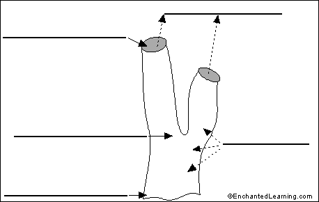
Label the letters on the diagrams found at www.biologycorner.com. Primordialis simple sponge (ascetta primordialis). And the simple act of parts of a sponge breaking of and establishing in a new location. However, sponges exhibit a range of diversity in body forms, including variations in the size of the spongocoel, the number of osculi, and where the cells that filter food from the water are located. Sponges, the members of the phylum porifera (/ p ə ˈ r ɪ f ər ə /; Draw and label a cross section of a typical sponge. Be sure to use the following terms in your description: Using a hand lens.observe and draw the sponge. Filtered water is then expelled through fewer, larger exhalant openings (oscules). A bud separating from the parent sponge and creating a new sponge elsewhere; Hydra is a coelenterate, along with the jellyfish (scyphozoa), sea. Label sponge external anatomy diagram using the definitions listed below, label the sponge and the flow of water through it. Be sure to use the following terms in your description:
Be sure to use the following terms in your description: Meaning 'pore bearer'), are a basal animal clade as a sister of the diploblasts. (beat up and draw water up into the sponge). The growth of stolons that develop into new individuals; Place the sample on a microscope slide, and a few drops of water and cover it with a coverslip.
Label Sponge External Anatomy - EnchantedLearning.com from www.enchantedlearning.com Spongin, spicules, ostia, choanocytes, osculum. Sponges, the members of the phylum porifera are a basal animal clade as a sister of the. Using a hand lens.observe and draw the sponge. Observe the sponge under the compound scope. Draw and label each of the specimens provided. How to draw hydrahow to draw hydra, how to draw hydraulic circuit diagram, how to draw hydra step by step, how to draw hydra easy, how to draw hydra in zoolo. Draw 5 ml of blood and discard before drawing the specimen tubes for testing. Draw and label a cross section of a typical sponge.
The sponges clipart gallery includes 48 illustrations of sponges.
Sponges, the members of the phylum porifera are a basal animal clade as a sister of the. The morphology and physiology of sponges were first adequately understood by who created in 1836 the name porifera for the group by which it is now generally known, iuxle (1875) and sollas (1884) proposed the complete separation of sponges from other metazoa on the grounds of many peculiarities. Hydra is a coelenterate, along with the jellyfish (scyphozoa), sea. Hermaphroditic, gamete, spawn, planktonic larvae. Include in your drawing the flow of water (and food particles) throughout the sponge in the process of filter feeding. Sponges, also called poriferans, are in the phylum porifera and are all sessile animals that live and feed attached to the bottom of the sea. Draw 5 ml of blood and discard before drawing the specimen tubes for testing. Label the collection tubes at the bedside or drawing area. Use the terms in the accompanying list to label the diagram,. Label sponge external anatomy diagram using the definitions listed below, label the sponge and the flow of water through it. (beat up and draw water up into the sponge). Using a hand lens.observe and draw the sponge. Draw a sea sponge and label its parts.
There are at least 5,000 named species of sponges, likely with thousands more yet to be classified. Write down 2 unique characteristics of each of the 4 different sponge types (encrusting, tubular, pectin, boring). Next, remove a tiny piece of your grantia sponge from the sample. Draw 5 ml of blood and discard before drawing the specimen tubes for testing. Spongin, spicules, ostia, choanocytes, osculum.
Overview of Sponges from web.augsburg.edu The sponges clipart gallery includes 48 illustrations of sponges. The instructions included specific information about sponge anatomy to be colored on diagram. Draw and label each of the specimens provided. The thin, flattened cells of the epidermis are called pinacocytes. Explain how a stinging cell in a cnidarian fires. Having observed the sponge anatomy, draw a simple sketch of a sponge and label the following parts: A collagenous matrix (the mesohyl) ills the space between the canals and chambers, harbouring Simple worksheet for labeling the parts of the sponge such as the osculum and choanocyte.
Although (1816) separated the sponges in a group spongiaria allied to protozoa.
The difference between this and the mechanisms of other animals. The sponge is a primitive aquatic vertebrate with a soft porous body supported by a framework of either fibers, calcareous, or spicules. The thin, flattened cells of the epidermis are called pinacocytes. Using a hand lens.observe and draw the sponge. The morphology and physiology of sponges were first adequately understood by who created in 1836 the name porifera for the group by which it is now generally known, iuxle (1875) and sollas (1884) proposed the complete separation of sponges from other metazoa on the grounds of many peculiarities. Digestion of the food particle takes place inside the cell. The instructions included specific information about sponge anatomy to be colored on diagram. Although (1816) separated the sponges in a group spongiaria allied to protozoa. Write down 2 unique characteristics of each of the 4 different sponge types (encrusting, tubular, pectin, boring). Make an illustration of your observations. How to draw hydrahow to draw hydra, how to draw hydraulic circuit diagram, how to draw hydra step by step, how to draw hydra easy, how to draw hydra in zoolo. The sponge's (a) basic body plan is a cylinder shape with a large central cavity. Explain the process of reproduction in the sponge: